MRI for Chronic Pain: When X-Rays and Ultrasounds Are Not Enough
MRI for Chronic Pain in Turkey helps doctors investigate persistent back, joint, muscle, and nerve pain when X-rays or ultrasound examinations do not provide enough information. MRI creates detailed images of soft tissues, spinal discs, cartilage, and nerves, helping identify certain conditions that conventional imaging may miss. For international patients, MRI Scan in Turkey offers a diagnostic option when a specialist determines that advanced imaging is necessary. However, MRI does not identify every cause of chronic pain, and doctors must interpret the findings alongside symptoms and clinical examinations. Living with persistent pain can affect sleep, mobility, work, and everyday activities. When initial examinations appear normal, patients may feel uncertain about the cause of their symptoms. Understanding when MRI is useful can help patients make informed decisions and avoid unnecessary investigations.


Understanding Chronic Pain
Chronic pain generally refers to pain that persists or recurs for more than three months. It may develop after an injury, surgery, or illness, or appear gradually without an obvious cause.
Unlike acute pain, chronic pain can involve changes in how the nervous system processes pain signals. This means the intensity of pain does not always correspond directly to visible tissue damage.
Common forms include persistent back pain, neck discomfort, joint pain, and nerve-related symptoms. Chronic pain can also affect concentration, emotional well-being, and quality of life.
Diagnosing the underlying cause often requires a combination of medical history, physical examination, and appropriate diagnostic tests.
A normal X-ray does not necessarily exclude a medical condition. However, not every cause of chronic pain produces abnormalities that imaging can detect.
Why X-Rays and Ultrasounds Are Often Not Enough
X-rays, ultrasound, and MRI provide different types of medical information.
X-rays are particularly useful for examining bones, fractures, joint alignment, and certain degenerative changes. However, they provide limited information about spinal discs, nerves, and many soft-tissue structures.
Ultrasound helps doctors examine muscles, tendons, ligaments, superficial nerves, and certain joints. It can also guide injections and assess some structures during movement.
However, ultrasound has limitations when examining deep tissues or structures hidden behind bone.
MRI for chronic pain provides detailed images of internal structures, particularly soft tissues. It may reveal abnormalities that are difficult to evaluate through other imaging methods.
Doctors may recommend MRI when symptoms persist, neurological findings raise concerns, or the results could influence treatment decisions.
However, a normal X-ray or ultrasound does not automatically mean an MRI is necessary.
How MRI for Chronic Pain Works
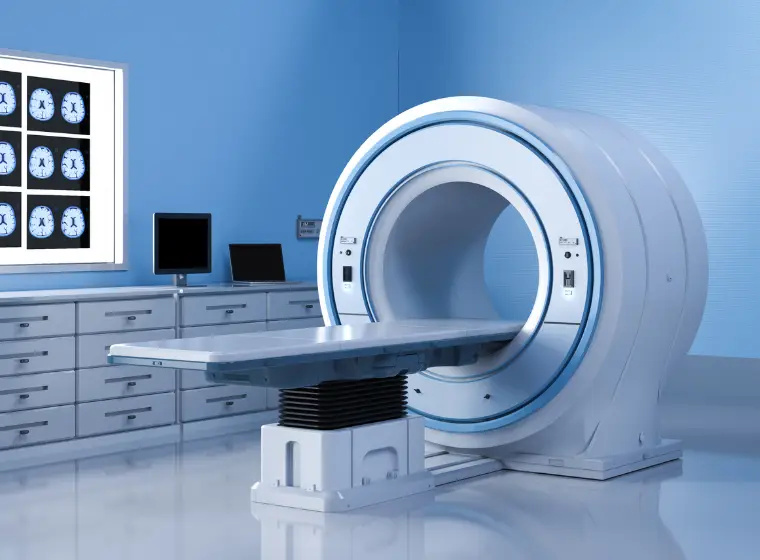
MRI uses a powerful magnetic field and radiofrequency pulses to create detailed images of the body's internal structures.
Unlike X-rays and CT scans, MRI does not use ionizing radiation.
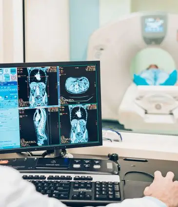
With MRI for chronic pain, doctors can examine spinal discs, ligaments, cartilage, muscles, bone marrow, and certain nerve structures.
Depending on the area being examined, MRI may help identify disc herniation, nerve compression, inflammation, ligament injuries, or other structural abnormalities.
However, imaging findings must always be interpreted alongside symptoms and physical examination results.
For example, disc degeneration and other age-related changes frequently appear on MRI scans in people who experience no pain.
An abnormal MRI finding does not automatically explain chronic pain, and a normal MRI does not exclude every possible cause.
Conditions MRI for Chronic Pain Can Detect
MRI can help investigate several conditions associated with persistent pain.
Common findings include:
- Herniated discs: Disc material may press against nearby nerves and contribute to pain or neurological symptoms.
- Spinal stenosis: Narrowing of the spinal canal or nerve openings may cause pain, numbness, or weakness.
- Ligament and tendon injuries: MRI can identify certain tears and soft-tissue abnormalities.
- Cartilage damage: Joint MRI may reveal cartilage defects or other internal joint changes.
- Inflammatory conditions: MRI can detect some forms of inflammation involving joints, bone marrow, or surrounding tissues.
- Occult fractures: Certain fractures or stress injuries may not appear on initial X-rays.
Although these findings can help guide diagnosis, their clinical significance depends on the patient's symptoms.
For individuals considering MRI Scan in Turkey, specialist evaluation helps determine which anatomical area requires imaging and whether the results are likely to influence treatment.
MRI for Different Types of Chronic Pain
The usefulness of MRI depends on the location and characteristics of the pain.
Back and Neck Pain
Spinal MRI can assess discs, the spinal canal, and nearby nerve structures. It may be appropriate when patients experience persistent nerve-related symptoms, progressive neurological problems, or other concerning findings.
However, routine MRI is generally not recommended for uncomplicated back pain without warning signs.
Knee, Hip, and Shoulder Pain
Joint MRI can help identify cartilage injuries, tendon tears, ligament damage, and certain abnormalities that may not appear on X-rays.
Doctors usually consider MRI when symptoms and examination findings suggest a condition requiring more detailed assessment.
Nerve-Related Pain
MRI may identify structural causes of nerve compression, particularly in the spine.
However, some nerve disorders do not produce visible MRI abnormalities. Additional tests, such as nerve conduction studies or electromyography, may be appropriate in selected cases.
The goal is to select the examination most likely to answer the clinical question rather than automatically choosing the most advanced imaging method.
What to Expect During an MRI
An MRI examination usually takes approximately 20–45 minutes, although the duration varies depending on the body part and imaging protocol.
During the scan, patients lie on a table that moves into the MRI scanner. Remaining still helps produce clear images.
The procedure itself is generally painless, although lying in one position may cause discomfort for people with chronic pain.
MRI scanners produce loud sounds, so patients usually receive hearing protection. People who experience claustrophobia should inform the medical team before the examination.
Before the scan, healthcare professionals check for implanted medical devices, metal fragments, and other potential safety concerns.
Some examinations require a gadolinium-based contrast agent, particularly when doctors need additional information about inflammation, tumors, infection, or certain postoperative changes.
Contrast is not necessary for most routine musculoskeletal MRI examinations. Patients should inform their healthcare team about relevant medical conditions, including kidney disease, before contrast administration.
How MRI for Chronic Pain Helps Treatment Decisions
One of the main advantages of MRI for chronic pain is its ability to provide detailed anatomical information when doctors suspect a structural condition.
For example, MRI findings may help specialists determine whether persistent symptoms are associated with nerve compression, ligament damage, or certain joint abnormalities.
This information can influence decisions about physical therapy, medication, targeted procedures, or surgery.
However, MRI does not automatically identify the source of pain or guarantee faster recovery.
Some abnormalities are incidental findings that do not require treatment. Doctors therefore compare MRI results with symptoms, medical history, and examination findings before recommending interventions.
When imaging does not reveal a clear structural cause, other approaches to chronic pain management may still provide meaningful relief.
Costs of MRI for Chronic Pain in Turkey
The cost of MRI for Chronic Pain in Turkey depends on the area being examined, the imaging protocol, and whether contrast is required.
A routine knee MRI may involve different technical requirements from a spinal MRI or an examination covering multiple anatomical regions.
Other factors may include radiologist reporting, specialist consultations, and additional diagnostic tests.
For international patients, MRI Scan in Turkey may form part of a broader diagnostic evaluation when a healthcare professional identifies a clinical need.
A general Check-up in Turkey can help review overall health concerns, but it does not automatically include MRI or replace a focused assessment of persistent pain.
Before arranging an examination, patients should clarify which body region requires imaging, whether contrast is necessary, and how the findings will be reviewed.
Choosing the appropriate examination is more important than undergoing multiple scans without a clear medical indication.
Final Thoughts
MRI for chronic pain can provide valuable diagnostic information when X-rays, ultrasound, and clinical examination do not adequately explain persistent symptoms.
It is particularly useful for evaluating certain spinal, neurological, joint, and soft-tissue conditions. However, MRI findings do not always identify the cause of pain, and advanced imaging is not necessary for every patient.al, joint, and soft-tissue conditions. However, MRI findings do not always identify the cause of pain, and advanced imaging is not necessary for every patient.
For individuals considering MRI Scan in Turkey, a focused medical assessment helps determine whether imaging is appropriate and how the findings may guide treatment.
Understanding the strengths and limitations of MRI supports more informed decisions about chronic pain diagnosis and management.
Frequently Asked Questions
1. Can MRI detect chronic pain that does not appear on X-rays?
MRI can identify certain soft-tissue, spinal, and joint abnormalities that X-rays cannot clearly show. However, not every cause of chronic pain produces visible changes on MRI.
2. Is MRI better than ultrasound for chronic pain?
Neither examination is universally better. MRI is useful for evaluating deep structures and certain spinal or joint conditions, while ultrasound is effective for many superficial soft-tissue problems. The appropriate test depends on the suspected condition.
3. Can an MRI be normal even when someone has chronic pain?
Yes. Chronic pain can occur without visible structural abnormalities. A normal MRI does not mean the pain is imaginary or unimportant.
4. Does every patient with chronic back pain need an MRI?
No. Doctors generally recommend MRI when symptoms, examination findings, or warning signs suggest that advanced imaging could influence diagnosis or treatment.
5. Is MRI included in a general medical check-up?
A general medical check-up does not automatically include MRI. Doctors usually recommend MRI separately when symptoms or clinical findings justify further investigation.
- Addiction ( 35 )
- Bariatric ( 7 )
- Cardiology ( 15 )
- Check-up ( 80 )
- Ear Nose & Throat ( 1 )
- Eye Care ( 4 )
- General Surgery ( 6 )
- Gynecology ( 7 )
- Hair Transplant ( 6 )
- Hematology ( 2 )
- IVF ( 22 )
- Longevity ( 27 )
- Neurosurgery ( 7 )
- Oncology ( 15 )
- Orthopedics ( 12 )
- Physiotherapy ( 1 )
- Plastic & Beauty ( 10 )
- Transplantation ( 4 )
- Urology ( 10 )