
Fractional Flow Reserve for Coronary Artery Disease
Fractional flow reserve measures whether a narrowed coronary artery is actually restricting blood flow, and the CT version does it without putting a wire or a catheter inside the heart. A blockage can look severe on an angiogram and still supply the heart perfectly well, while a modest-looking narrowing can cause real symptoms, and this test tells the two apart. It runs off a coronary CT angiography scan with computer modelling of pressure along the vessel, so there is no hospital stay and no recovery time, and patients arranging it in Turkey should check whether the CT scan itself is included in the fee. Coronary artery disease continues to prevail as one of the most common and dangerous heart conditions worldwide. Some individuals experience chest pain, shortness of breath, or fatigue without knowing that their heart arteries may be narrowing. Traditional tests like angiography or stress tests provide valuable information, but sometimes they do not accurately reflect the severity of the blockage. This is where CT fractional flow reserve comes in, an advanced method that provides doctors with a clearer, more accurate picture of how well blood flows through the coronary arteries.

What Is CT Fractional Flow Reserve?
CT fractional flow reserve (also called FFR-CT) is a non-invasive technique used to determine whether a coronary artery blockage is actually restricting blood flow. Instead of inserting wires or tools into the heart, this technique uses high-resolution CT images and computer modelling to calculate pressure differences in the vessels. It shows how much blood can move through a narrowed artery and whether the narrowing is severe enough to require treatment. The idea is simple: a blockage may appear significant on imaging but not always reduce blood flow. CT fractional flow reserve helps distinguish harmless narrowings from those that pose a real risk to the heart.
How CT Fractional Flow Reserve Works
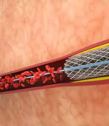
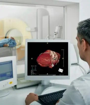
The process begins with a coronary CT angiography scan. This scan captures detailed images of the heart and its arteries. Using these images, advanced software creates a 3D model of the coronary vessels. It then simulates blood flow and measures the pressure at various points in the arteries. If the simulated pressure drops below a certain threshold, it means that blood flow is significantly reduced. This information helps cardiologists decide whether a patient needs medication, a stent, or further invasive testing. The entire process is painless and requires no hospital stay. It allows patients to continue their day without recovery time, a significant advantage of CT fractional flow reserve.
Why CT Fractional Flow Reserve Is More Accurate
Traditional angiography can sometimes be misleading because it only shows the shape of the artery, not how well blood flows through it. A vessel might appear severely narrowed yet still allow sufficient blood flow. In other cases, a seemingly minor narrowing can cause severe symptoms. CT fractional flow reserve provides a functional assessment; it shows what is happening with the blood flow in real time. By combining anatomical imaging with physiological data, FFR-CT offers a more complete understanding of coronary artery disease.
Benefits for Patients
There are several reasons why CT fractional flow reserve has become a preferred tool in modern cardiology:
- Completely non-invasive: No wires, catheters, or needles inside the heart.
- Accurate diagnosis: Indicates whether a blockage is dangerous.
- Avoids unnecessary procedures: Helps prevent unneeded stents or invasive angiography.
- Improves decision-making: Doctors can choose the best treatment with confidence.
- Fast results: Patients receive clear explanations without extended hospital stays.
Overall, FFR-CT offers peace of mind by showing what truly matters: how well the heart is being supplied with blood.
When Doctors Recommend CT Fractional Flow Reserve
Doctors may suggest CT fractional flow reserve in several situations:
- When a coronary CT angiogram shows a blockage of uncertain severity.
- When a patient has chest pain, but test results are inconclusive.
- Before recommending a stent procedure.
- When patients prefer a non-invasive option before undergoing catheter-based testing.
Why CT Fractional Flow Reserve Is Growing in Turkey
Turkey has become a leading destination for modern heart diagnostics, including FFR-CT. Hospitals in major cities use advanced CT scanners and specialized software that meet international standards. Patients benefit from experienced cardiologists, short waiting times, and streamlined patient care.
Costs of CT Fractional Flow Reserve in Turkey
The cost of FFR-CT varies by hospital, the imaging technology used, and whether additional tests are needed. Even so, Turkey remains more affordable compared with many European and Western countries, while offering high-quality care and advanced heart imaging.
Final Thoughts
CT fractional flow reserve is transforming the way doctors diagnose and manage coronary artery disease. It offers a safe, non-invasive, and highly accurate way to understand whether a blockage is truly harmful. For patients seeking clarity, confidence, and modern heart care, FFR-CT offers a robust, reliable solution.
Frequently Asked Questions About Fractional Flow Reserve
1. What is the difference between FFR-CT and invasive FFR?
Invasive FFR measures pressure with a thin wire threaded into the coronary artery during a catheter procedure. FFR-CT reaches the same kind of answer by modelling blood flow from a CT scan, with nothing entering the body. The invasive version is still used when a catheter procedure is happening anyway or when the CT result is unclear.
2. Do I need a separate scan for the test?
The measurement is calculated from a coronary CT angiography scan, so that scan has to be done first. If you have already had a recent, good-quality coronary CT, the images can sometimes be used without repeating it. Otherwise the CT and the FFR analysis are handled as one appointment.
3. Does the test hurt or need a hospital stay?
No. You lie on the CT table for the scan, which involves a contrast injection through a vein in the arm, and the analysis happens afterwards on a computer. There is no sedation and no observation period, so you can go about your day once the scan is finished.
4. Can FFR-CT replace an invasive angiogram completely?
For many patients it can, because a normal result means an artery is unlikely to need a stent and the catheter procedure can be avoided. It does not replace angiography when treatment is clearly needed, since a stent still has to be placed through a catheter. Its main value is filtering out the people who would have had an invasive test for nothing.
5. Are there patients the test is not suitable for?
The analysis depends on clear CT images, so heavy calcification, an irregular heart rhythm or previous stents in the vessel of interest can make the result unreliable. Contrast allergy and reduced kidney function also need to be considered before any CT with contrast. A cardiologist will weigh these up against the alternatives before recommending it.
- Addiction ( 34 )
- Bariatric ( 7 )
- Cardiology ( 15 )
- Check-up ( 74 )
- Ear Nose & Throat ( 1 )
- Eye Care ( 4 )
- General Surgery ( 6 )
- Gynecology ( 7 )
- Hair Transplant ( 6 )
- Hematology ( 2 )
- IVF ( 22 )
- Longevity ( 25 )
- Neurosurgery ( 7 )
- Oncology ( 15 )
- Orthopedics ( 12 )
- Physiotherapy ( 1 )
- Plastic & Beauty ( 10 )
- Transplantation ( 4 )
- Urology ( 10 )